Home
/ Cranial Nerves Diagram Labeled : Cranial Nerves I Ii I Olfactory Sensory Smell Ppt Video Online Download - Human anatomy and physiology weakness in the sixth cranial nerve can appear within a few hours or days after vaccination.
Cranial Nerves Diagram Labeled : Cranial Nerves I Ii I Olfactory Sensory Smell Ppt Video Online Download - Human anatomy and physiology weakness in the sixth cranial nerve can appear within a few hours or days after vaccination.
Cranial Nerves Diagram Labeled : Cranial Nerves I Ii I Olfactory Sensory Smell Ppt Video Online Download - Human anatomy and physiology weakness in the sixth cranial nerve can appear within a few hours or days after vaccination.. Labeled diagram with brain sections. All cranial nerves originate from nuclei in the brain.two originate from the forebrain (olfactory and optic), one has a nucleus in the spinal cord (accessory) while the. The cranial nerves are loosely based on their functions. But what are their names and functions? The cranial nerves (cn) are twelve pairs of nerves that, with the exception of the spinal accessory nerve (cn xi), originate in the brain and contribute to the peripheral nervous system (pns), supplying the head and neck.
Cranial nerve anatomy / cranial nerves. Learn vocabulary, terms, and more with flashcards, games, and other study tools. Let's look at that now. Start studying anatomy and physiology brain and cranial nerves. The cranial nerves are all located on the underside of your brain inside your skull.
What Are Cranial Nerves How Many Cranial Nerves Are There from www.scienceabc.com The cranial nerves are all located on the underside of your brain inside your skull. Aspects of vision, like peripheral vision, are under the control of the optic cranial nerve (ii). Start studying cranial nerves labeling practice (brain). They come in pairs, one on each side of the brain, and are numbered in roman numerals i through xii. Cranial nerve anatomy / cranial nerves. This human anatomy module is about the cranial nerves. Let's look at that now. Your cranial nerves are pairs of nerves that connect your brain to different parts of your head, neck, and trunk.
Controls the sternocleidomastoid and trapezius muscles, and overlaps with functions of the vagus nerve (cn x).
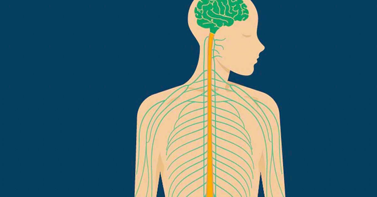
All the nerves are distributed in the head and neck except the tenth, which also supplies structures in the thorax and abdomen. Human anatomy and physiology weakness in the sixth cranial nerve can appear within a few hours or days after vaccination. Spinal nerves emerge sequentially from the spinal cord with the spinal nerve closest to the head (c1) emerging in the space above the first cervical vertebra. The cranial nerves are part of the peripheral nervous system this diagram labels the cranial nerves. This nerve performs two major functions. Learn vocabulary, terms, and more with flashcards, games, and other study tools. All cranial nerves originate from nuclei in the brain.two originate from the forebrain (olfactory and optic), one has a nucleus in the spinal cord (accessory) while the. The cranial nerves are loosely based on their functions. Start studying anatomy lab practical 1: The cranial nerves are all located on the underside of your brain inside your skull. Diagram | dinah (marc schneider) each has a different function for sense or this article will explore the functions of the cranial nerves and provide a diagram. This mri cranial nerves axial cross sectional anatomy tool is absolutely free to use. They come in pairs, one on each side of the brain, and are numbered in roman numerals i through xii.
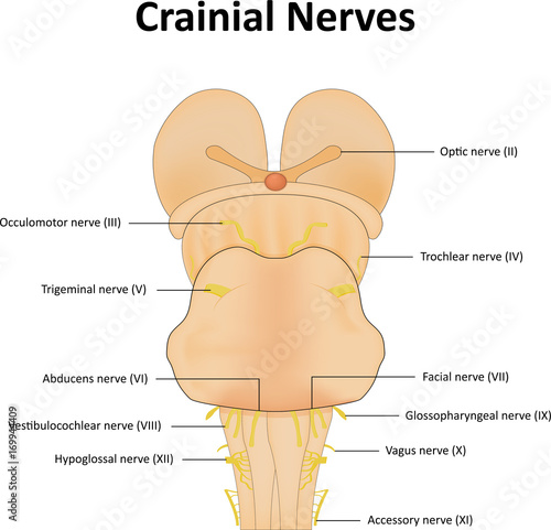
Learn vocabulary, terms, and more with flashcards, games, and other study tools. (a) schematic diagram showing the nuclei within the brainstem involved in various cranial nerves, with depiction of the efferent (motor) nuclei on one side and afferent (sensory) nuclei on the other. The cranial nerves (origin, pathways & applied anatomy) there are twelve cranial nerves, which leave the brain and pass through foramina in the skull. This diagram depicts cranial nerves anatomy.human anatomy diagrams show internal organs, cells, systems, conditions, symptoms and sickness information and/or tips for healthy living. There are 12 of them, each named for their function or structure.
Cranial Nerves Labeled Diagram Stock Vector Adobe Stock from as1.ftcdn.net Each nerve has a name that reflects its function and a number according to its location in the brain. Cranial nerve anatomy / cranial nerves. The cranial nerves emerge from the central nervous system above this level. Each cranial nerve is paired and is present on both sides. The added annotations detail their attachment to the brain, their cranial exit, innervations, functions and nerve type. Start studying anatomy and physiology brain and cranial nerves. Mainly motor cranial and spinal roots located in the jugular foramen. Let's look at that now.
This nerve performs two major functions.
Cranial nerve anatomy / cranial nerves. Cranial nerve anatomy and terminology. Start studying anatomy lab practical 1: Start studying anatomy and physiology brain and cranial nerves. This human anatomy module is about the cranial nerves. Simple line diagrams accompany the text. Unit 1 biology basics bio b. (a) schematic diagram showing the nuclei within the brainstem involved in various cranial nerves, with depiction of the efferent (motor) nuclei on one side and afferent (sensory) nuclei on the other. The cranial nerves (cn) are twelve pairs of nerves that, with the exception of the spinal accessory nerve (cn xi), originate in the brain and contribute to the peripheral nervous system (pns), supplying the head and neck. They come in pairs, one on each side of the brain, and are numbered in roman numerals i through xii. The cranial nerves are twelve pairs of nerves from the central nervous system. The cranial nerves emerge from the central nervous system above this level. The cranial nerves are all located on the underside of your brain inside your skull.
The cranial nerves are loosely based on their functions. Simple line diagrams accompany the text. These are often labeled as cn i, cn ii, and so on. Cranial nerve anatomy and terminology. In this summary, we discuss the nomenclature of the cranial nerves and supply some background information that might make it easier to understand the nerves and their function.
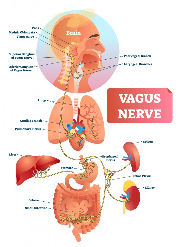
12 Cranial Nerves Nerves Functions Diagram Of Locations from post.healthline.com Controls the sternocleidomastoid and trapezius muscles, and overlaps with functions of the vagus nerve (cn x). Illustration about central, brain, head, human, gland, external, body, lungs, labeled. The cranial nerves comprise 12 nerves of the peripheral nervous system which originate from brain nuclei and exit from the foramina and fissures of the cranium. Learn vocabulary, terms, and more with flashcards, games, and other study tools. The chapter begins with a brief review of the anatomy of the skull base. The cranial nerves (cn) are twelve pairs of nerves that, with the exception of the spinal accessory nerve (cn xi), originate in the brain and contribute to the peripheral nervous system (pns), supplying the head and neck. Spinal nerves emerge sequentially from the spinal cord with the spinal nerve closest to the head (c1) emerging in the space above the first cervical vertebra. Start studying anatomy and physiology brain and cranial nerves.
Your cranial nerves are pairs of nerves that connect your brain to different parts of your head, neck, and trunk.
Each cranial nerve is paired and is present on both sides. This is an online quiz called cranial nerves label. There are 12 paired cranial nerves that arise from the brainstem. Learn vocabulary, terms, and more with flashcards, games, and other study tools. Start studying cranial nerves labeling practice (brain). Learn vocabulary, terms, and more with flashcards, games, and other study tools. The cranial nerves are named as follows; Cranial nerves model labeled biol 160: Game to label the 12 cranial nerves and other visible structures. Learn vocabulary, terms, and more with flashcards, games, and other study tools. Your cranial nerves are pairs of nerves that connect your brain to different parts of your head, neck, and trunk. Simple line diagrams accompany the text. It conveys some sensory information from the tongue and the interior of the mouth.